Posts

Kidney Anatomy

Human kidneys are paired, reddish brown, bean shapped structures about 11 cm (4.4 in) long and function as excretory system . They are located in back of the body cavity, one on each side of the spine just above the waist. The kidneys are loosely held in place by a mass of fat and by fibrous tissue. The outer margin is convex, the inner border concave. On the inner surface is a slit, the hilus, through which pass the arteries, veins, nerves and the renal pelvis, a funnellike structure. Urine from each kidney is collected in the renal pelvis and passes into the hollow tube, the ureter, which extends downward, emptying into the urinary bladder. A shorter, single tube, the urethra, eliminates urine the bladder. The cut surface of the kidney reveals two distinct areas; the cortex - a dark band along the outer border, about 1 cm (0.4 in) thickness, and the inner medulla. The medulla is divided into 8 to 18 conical tissues termed renal pyramids (cortical arches) and extends down ...
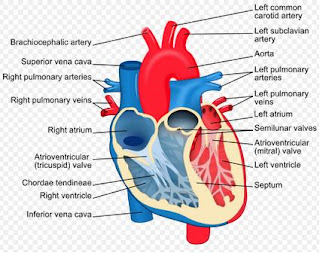
Heart

Image
The human heart is a specialized, have four chambered muscle that maintains blood flow in the circulatory system. The heart is located in the thorax, it lies on the left of the body's mid-line, above and in contact with the diaphragm. It is situated immediately behind the breastbone, or sternum, and between the lungs, with its apex titled to the left. At rest the heart pumps about 59 cc (2 oz) of blood per beat and 5 l (5 qt) per minute, compared to 120 - 220 cc (4-7.3 oz) per beat and 20-30 l (21-32 qt) per minute during exercise. The adult human heart is about 250 - 350 gram (9 oz). Blood supplies food and oxygen to the cells of the body for their life needs and removes the waste products of their chemical processes. It also helps to maintain a consistent body temperature, circulate hormones, and fight infections. Research indicates that the heart itself produces a hormonelike chemical. Brain cells are dependent on a constant oxygen supply, so death ensues shortly...

Spinal Cord

Together with the brain, the spinal cord constitutes an important part of the central nervous system. It conducts impulses between the brain and the rest of the body and is a center for reflex activity. The spinal cord is present in the vertebral column of all vertebrate animals, running from the brain stem to the tail. The spinal cord consists of a tube about 1 cm thick with a small central canal. Cerebrospinal fluid flow through this canal and around the spinal cord. The cord is encased in three membranes called meninges; the pia mater, the arachnoid, and the dura mater. In cross section, the spinal cord has centrally positioned butterfly shaped gray matter, consisting of unmyelinated cell bodies of nerve cell (neurons), surrounded by the myellinated axonal fibers, or white matter. A cell body contains the nucleus of the nerve cell, and it sends out a single axon and numerous fibrous extensions known as dendrites. In the spinal cord, dendrites receive stimuli from the ax...

Breast

Breast, in human females, are two glandular organs that secret milk for feeding newborn infants. Such gland are present in all mammals and are known in general as mammaries. Physiologycally, they are highly modified sweet glands. No other animals posses such glands, and no related precedents exist in other classes of vertebrates. Rudimentary breasts are present in both human sexes at birth. No further development takes place in males during normal growth, but full development occurs in females in the early childbearing period. A projection called a nipple is found at the tip of each breast in both men and women. It is surrounded by a pigmented areas, the areola, that enlarges and deepens in color on pregnancy. Structure The female human breast consists mainly of a round mass of glandular tissue comprising about 15-20 lobes, each having a duct leading to an opening on the nipple, the duct system and glandular tissue fully develop with pregnancy. The amount of fat sheathing the glan...

Bone

Bone is a type of skeletal tissue. Bones are also organs made largely of this tissue and surrounded by a membrane called the periosteum. This study of bones is called osteology. Bone tissue contains living cells embedded in a hard matrix. This matrix consists mostly of calcium phosphate (as hydroxylapatite) and other calcium minerals held together by collagen (a protein) and other organic substances. The major types of bone tissue exist. Compact bone tissue has many rodlike Haversian systems, each containing many concentric cylindrical layers (lamellae) surrounding a central blood vessel . Between these layers the bone cells (osteocytes) lie within spaces called acunae. Tiny pores (canalculi) connect the osteocytes with one another and with the Blood vessel, allowing oxygen and nutrients to reach the cells. Cancellous, or spongy bone tissue has a bone network of rigid beams (trabeculae). The spaces between these  trabeculae contain marrow, a blood forming tissue. Young marrow...

Peristalsis

Peristaltis is one of several types of patterns of movement associated with the gastrointestinal tract. It can be described as a moving, coordinated wave of muscular contraction that is propulsive in nature, forcing the contents of the digestive tract ahead of it. Peristalsis occurs most frequently in the esophagus, where it is stimulated by a swallow (primary peristalsis) and moves the entire length of the esophagus to the stomach . A peristalsis wave may also occur in the absence of a swallow (secondary peristalsis). A secondary peristalsis is elicited when the esophagus is distended - if the primary wave fails to clear the esophagus of food, or if gastric contents reflux into the esophagus. Peristaltic waves also occur in the small intestine but rarely travel more than 4 cm (1.6 in). In the large intestine peristalsis is responsible for the two or three mass movements that occur daily. Peristalsis is controlled largely by central and local serves.

Stomach

Image
Stomach is part of the digestive system and receives food from the esophagus. A bottle shaped organ in humans. It is normally less than 20 cm (18 in) long and weighs about 1 percent of the body weight. It is capable, however, of distending greatly to accommodate large meals without increasing the pressure to tension on its walls. The stomach secretes large amounts of hydrochloric acid, which digests protein, activates pepsinogen to form the enzyme pepsin, and kills bacteria that enter the stomach. Acid is secreted from the oxyntic gland area of the gastric mucosa; other cells in this area secrete mucus and the proenzyme pepsinogen. Mucosa lubricates and protects the stomach wall and prevents acid from digesting the stomach itself. Pepsin is an enzyme that aids in the digestion of protein . The mucosa of the distal third of the stomach (antrum) produces and releases the hormone gastrin into the bloodstream), which stimulates acid secretion and the growth of mucosa in the digestiv...